
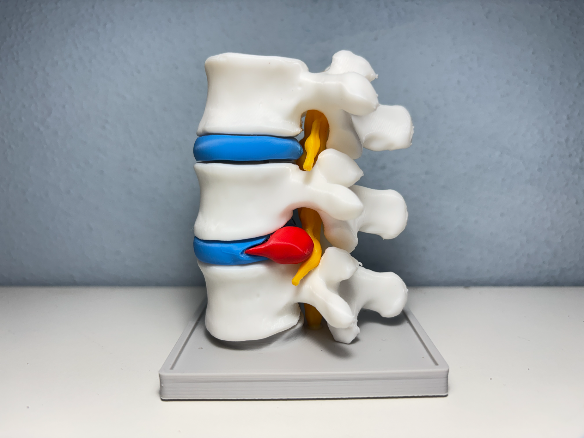
(NO AMS) Herniated Disc - Lumbar Spine
Peso
91g
Tiempo
6h 32m
Precio
Cotiza este producto por WhatsApp
Contáctanos y te damos el precio según color y acabado
Las dimensiones dependen del diseño. Confirmamos las medidas exactas por WhatsApp.
Descripción
After working on several complex and time-consuming anatomical models, I wanted to design something accessible to everyone. This Lumbar Spine with Herniated Disc requires no AMS and prints quick. Hope you find it useful for your desk, clinic, or classroom! Follow the simple video to assemble it. Since the order of the vertabrae could be misunderstood, they are clearly named in the print profile. Moreover, they are positioned on the plate from LEFT (bottom vertebra) to RIGHT (up vertebra). 🩻 Overview & Clinical Context This high-precision 3D anatomical model represents a section of the lumbar spine showcasing an intervertebral disc herniation. The model visually demonstrates the mechanics of nerve root compression: the focal protrusion of disc material (nucleus pulposus) into the spinal canal, directly impinging on the exiting nerve root. It serves as an intuitive tool for explaining the origin of common symptoms such as lower back pain, sciatica, and paresthesia. 🧠 Anatomy Explained (In Simple Terms) To make this model an effective educational tool for everyone—even without a medical background—here is a simple breakdown of what each part represents: 🦴 Lumbar Vertebrae (L3, L4, L5): The "L" stands for Lumbar, which refers to the lower back. These three heavy-duty bones carry most of your body weight and stack together to form the main structural column of your lower spine. 🔵 Intervertebral Discs (The Shock Absorbers): Positioned between each bone, these discs act like rubbery cushions or jelly-filled pads. They prevent the bones from rubbing against each other and allow your back to bend, twist, and absorb impact. 🔴 Herniated Disc (The Squeezed Gel): When an intervertebral disc is damaged, its soft gel-like interior bulges out through a tear in the outer wall. In this model, the red protrusion highlights an active herniation breaking out of its normal boundary. 🟡 Spinal Nerves & Sciatica: The yellow branches extending from the sides are spinal nerves that travel down into your legs. As shown, when the hernia pushes to the side (Posterolateral Herniation), it pinches a single nerve root, causing typical Sciatica—sharp, radiating pain or numbness down one leg. 🐴 Cauda Equina (The Central Nerve Bundle): Meaning "Horse's Tail" in Latin, this central yellow core represents the collection of nerve roots at the bottom of the spinal cord. Printing Design Note: Anatomically, the cauda equina is a collection of delicate, individual nerve strands floating in spinal fluid. To ensure a durable, easy-to-print model that doesn't snap during post-processing or handling, this bundle has been consolidated into a single solid form. ⚠️ What if the hernia pushes straight back into the Cauda Equina? While this model illustrates a classic side-pushing (posterolateral) hernia compressing a single nerve, a central herniation behaves very differently: If the disc bulges straight backward into the spinal canal, it presses directly onto the entire Cauda Equina bundle instead of just one side nerve. This is a critical medical condition called Cauda Equina Syndrome. Unlike typical sciatica, direct central compression can cause severe symptoms on both legs simultaneously, numbness in the saddle area (inner thighs and groin), and a sudden loss of bowel or bladder control—requiring urgent emergency surgery. 🎯 Intended Use Patient Communication: Ideal for orthopedic, neurosurgical, chiropractic, and physical therapy clinics to visually explain the nature of disc pathologies to patients. Medical Education: A valuable tactile and visual aid for students in medicine, physical therapy, healthcare, and anatomy. 3D Printing Showcase: An engaging educational project highlighting the capabilities of 3D printing in medical visualization. 💡 Printing & Assembly Recommendations ❌ No AMS Required: Designed and split specifically to be 100% printable on any FDM 3D printer. 🧩 Organized across 5 Plates (Color-Separated): Files are pre-arranged into 5 distinct print plates for effortless filament management without material waste:Plate 1 (Red): Herniated disc Plate 2 (Yellow): Nerve roots and cauda equina block Plate 3 (Grey / Black): Display stand base Plate 4 (Blue): Intervertebral discs Plate 5 (Bone White): Lumbar vertebrae Recommended Material: Standard PLA for all components. ⚠️ Disclaimer: This model is intended strictly for educational, illustrative, and hobbyist purposes. It is not a clinical tool and does not replace professional medical diagnosis, advice, or treatment. 📚 Medical References & Further Reading For a detailed clinical breakdown of lumbar disc disease and herniation symptoms, check out the Johns Hopkins Medicine Health Guide.
Tags
Diseñador
IceGabrielito