
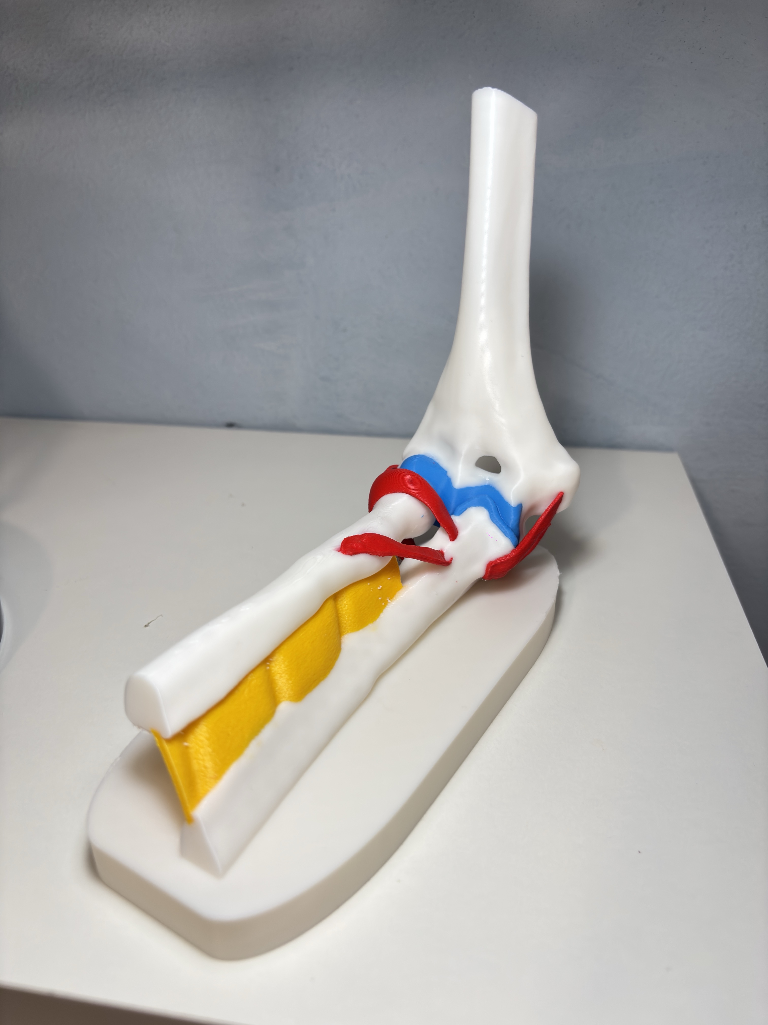
Detailed Elbow Joint Anatomy & Ligament Model
Peso
169g
Tiempo
8h 55m
Precio
Cotiza este producto por WhatsApp
Contáctanos y te damos el precio según color y acabado
Las dimensiones dependen del diseño. Confirmamos las medidas exactas por WhatsApp.
Descripción
Boost MeBoost for other anatomical models 🫀⚕️🩻 Overview 🩻 This highly detailed 3D model offers a precise, true-to-life representation of the human elbow joint. Meticulously designed using advanced anatomical references, it captures the complex biomechanical relationship between the bones and their stabilizing soft tissues. The model includes integrated display bases to showcase multiple angles, making it an exceptional, premium educational resource for medical students, physical therapists, biology teachers, and 3D printing enthusiasts looking for a striking anatomical piece. Included Anatomical Structures The model encompasses all essential components required to fully visualize the functional anatomy of the elbow: 🦴 Skeletal Framework: Humerus, Radius, and Ulna, featuring accurate anatomical landmarks and articulating surfaces. 🔗 Ligaments & Connective Tissues: Ulnar Collateral Ligament Complex, Radial Collateral Ligament, Lateral Ulnar Collateral Ligament, Annular Ligament of Radius, Oblique Cord, and the Interosseous Membrane. 🧬 Articular Surfaces: Articular Cartilage mapped precisely to the joint friction and load-bearing zones. Intended Use Academic & Educational: A perfect hands-on, tactile aid for university anatomy labs, medical schools, and healthcare training programs. Clinical Demonstration: Assists orthopedic specialists and physiotherapists in visually explaining joint mechanics or conditions (like ligament tears or "tennis elbow") directly to patients. Showpiece for Makers: An impressive, multi-material print that highlights the precise capabilities of modern 3D printers when handling complex organic shapes. 💡 Print Settings & Recommendations Material Compatibility: Engineered to be printed entirely in standard PLA. No TPU or flexible materials are needed, ensuring a straightforward and reliable printing experience on standard desktop machines. Multi-Color Ready (4 Colors): Fully optimized for a single 4-slot AMS/MMU system. To match the high-contrast educational layout seen in the photos, we suggest the following filament assignment: White: Skeletal framework (Humerus, Radius, Ulna) and bases Red: Ligaments and Oblique Cord Yellow: Interosseous Membrane Blue: Articular Cartilage Disclaimer: This 3D model is created exclusively for educational, illustrative, and hobbyist purposes. It is not intended for clinical use and should never substitute professional medical diagnosis, advice, or treatment.
Tags
Diseñador
IceGabrielito